43 parts of the eye without labels
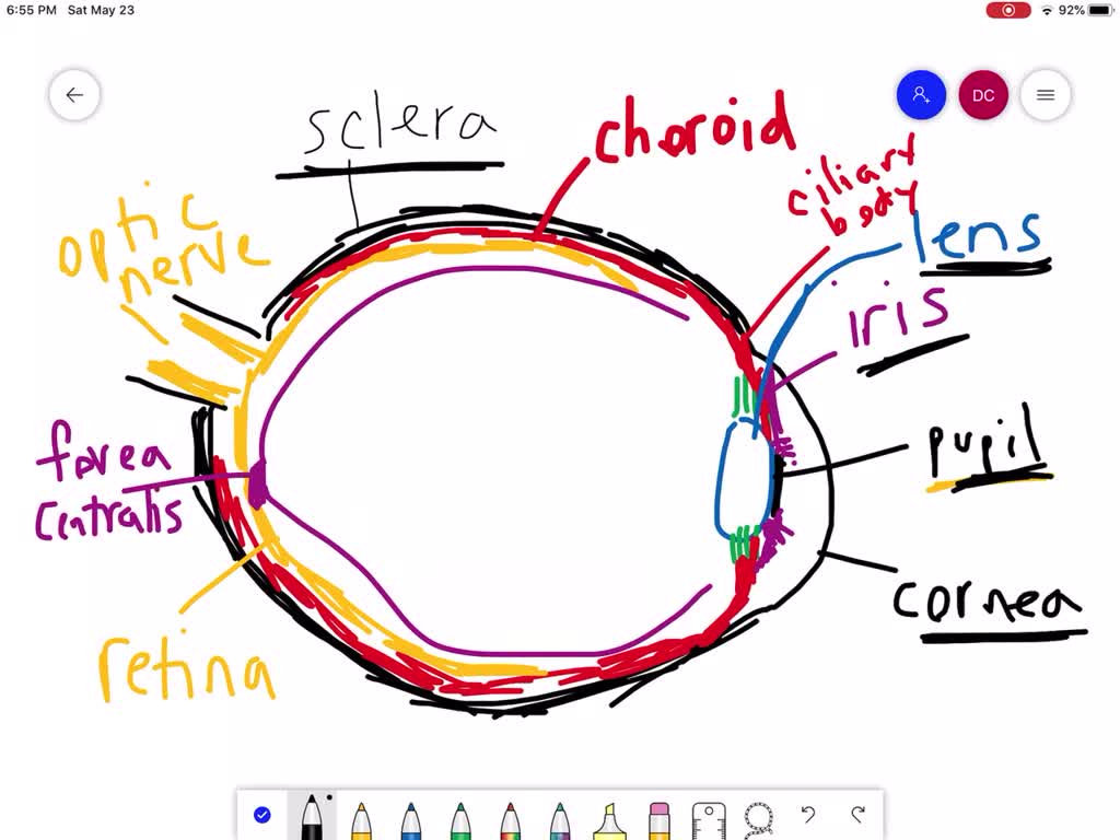
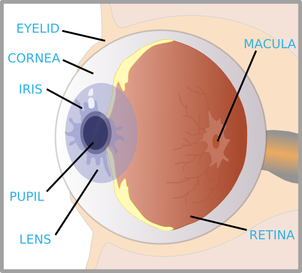
PDF Parts of the Eye Macula: The macula is the small, sensitive area of the retina that gives central vision. It is located in the center of the retina. Optic nerve: The optic nerve is the largest sensory nerve of the eye. It carries impulses for sight from the retina to the brain. Pupil: The pupil is the opening at the center of the iris. Eye Anatomy: 16 Parts of the Eye & Their Functions - Vision Center The following are parts of the human eyes and their functions: 1. Conjunctiva. The conjunctiva is the membrane covering the sclera (white portion of your eye). The conjunctiva also covers the interior of your eyelids. Conjunctivitis, often known as pink eye, occurs when this thin membrane becomes inflamed or swollen. Other eye disorders that affect the conjunctiva include:
PDF Eye Anatomy Handout - National Eye Institute Here are descriptions of some of the main parts of the eye: Cornea: The cornea is the clear outer part of the eye's focusing system located at the front of the eye. Iris: The iris is the colored part of the eye that regulates the amount of light entering the eye. Lens: The lens is a clear part of the eye behind the iris that helps to

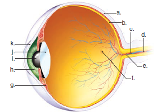
Parts of the eye without labels
The Eyes (Human Anatomy): Diagram, Optic Nerve, Iris, Cornea ... - WebMD Iris: the colored part. Cornea: a clear dome over the iris. Pupil: the black circular opening in the iris that lets light in. Sclera: the white of your eye. Conjunctiva: a thin layer of tissue ... Human Eye Ball Anatomy & Physiology Diagram - eMedicineHealth The eye is composed of various parts, all of which work together to allow the sight to occur. Cornea The cornea is the transparent, clear layer at the front and center of the eye. In fact, the cornea is so clear that one may not even realize it is there. The cornea is located just in front of the iris, which is the colored part of the eye. Eye Diagram With Labels and detailed description - BYJUS Cornea is a dome-shaped tissue covering the front of the eye. Iris is the coloured part of the eye and controls the amount of light entering the eye by regulating the size of the pupil. The lens is located just behind the iris. Its function is to focus the light on the retina. The optic nerve transmits electrical signals from the retina to the brain.
Parts of the eye without labels. rsscience.com › stereo-microscopeParts of Stereo Microscope (Dissecting microscope) - Rs' Science Optical parts of a stereo microscope work together to magnify and produce a 3-D image of the specimens. These parts include: Eyepieces. The eyepiece (or ocular lens) is the lens part at the top of a microscope that the viewer looks through. Typically, standard eyepieces for a dissecting microscope have a magnifying power of 10x. Parts of the Eye & Their Function | Robertson Optical and Optometry The iris is the area of the eye that contains the pigment which gives the eye its color. This area surrounds the pupil, and uses the dilator pupillae muscles to widen or close the pupil. This allows the eye to take in more or less light depending on how bright it is around you. If it is too bright, the iris will shrink the pupil so that they ... › engine-vu › yamaha-marine-genuineComplete Yamaha Marine Outboard OEM Parts Catalog | PartsVu Yamaha outboard parts are the heart and soul of PartsVu. Offering more than 40,000 Genuine Yamaha outboard parts and products and at low prices with same business day shipping on in-stock items. We value convenience and knowledge, providing resources like our Yamaha Outboard Maintenance Parts Charts , helping you to find the right parts every time. BYJUS BYJUS
leafyplace.com › parts-of-a-flowerParts of Flower and Plant (Pistil, Sepal, Stamen and More ... Dec 26, 2019 · Extracts of many plants have medicinal properties that can help address a number of ailments. Parts of plants can be used to make therapeutic herbal teas, essential oils, or taken as supplements. Pleasing to the eye. Flowers, blossoms, and colorful petals are pleasing to the eye. diagram of eye with labels Ear anatomy eye models lab guide structures identify biologycorner brain senses biology eyes physiology parts use answers. Peregrine diagram falcon hi res. Picture of the eye labeled elegant human eye anatomy for kids ... Eye Diagram Without Labels | Via Anatomy Pictures Gallery If… | Flickr . liver fluke. Perception: 3.1 Eye To ... Quiz: Label The Parts Of The Eye - ProProfs How much did you get to understand about the human eye? Take up this quiz and find out! Questions and Answers. 1. A is pointing to what part of the eye? A. Cornea. B. Optic Nerve. diagram of a label eye Eye Diagram Without Labels | via Anatomy Pictures Gallery if… | Flickr. 11 Pics about Eye Diagram Without Labels | via Anatomy Pictures Gallery if… | Flickr : The Eye and Vision, Pinguecula eye causes, symptoms, diagnosis & pinguecula eye treatment and also label the muscles of the eye - PurposeGames. ... eye human anatomy eyes parts ...
Anatomy of the eye: Quizzes and diagrams - Kenhub How to learn the parts of the eye. Found within two cavities in the skull known as the orbits, the eyes are surrounded by several supporting structures including muscles, vessels, and nerves. There are 7 bones of the orbit, two groups of muscles (intrinsic ocular and extraocular), three layers to the eyeball… and that's just the beginning. There's a lot to learn, but stay calm! Eye Anatomy Detail Picture Image on MedicineNet.com The eye is our organ of sight. The eye has a number of components which include but are not limited to the cornea, iris, pupil, lens, retina, macula, optic nerve, choroid and vitreous. Cornea: clear front window of the eye that transmits and focuses light into the eye. Iris: colored part of the eye that helps regulate the amount of light that enters Anatomy of the Eye. Learn about the different parts of the eye. The sclera is a membrane of tendon in the eye, also known as the white of the eye. Rugged and robust, the sclera works to protect the inner, more sensitive parts of the eye like the retina and choroid. It is about 0.03 of an inch thick except for where the four "straight" eye muscles append, where the depth is no more than 0.01 of an inch. Eye Diagram With Labels and detailed description - BYJUS Cornea is a dome-shaped tissue covering the front of the eye. Iris is the coloured part of the eye and controls the amount of light entering the eye by regulating the size of the pupil. The lens is located just behind the iris. Its function is to focus the light on the retina. The optic nerve transmits electrical signals from the retina to the brain.
Human Eye Ball Anatomy & Physiology Diagram - eMedicineHealth The eye is composed of various parts, all of which work together to allow the sight to occur. Cornea The cornea is the transparent, clear layer at the front and center of the eye. In fact, the cornea is so clear that one may not even realize it is there. The cornea is located just in front of the iris, which is the colored part of the eye.
The Eyes (Human Anatomy): Diagram, Optic Nerve, Iris, Cornea ... - WebMD Iris: the colored part. Cornea: a clear dome over the iris. Pupil: the black circular opening in the iris that lets light in. Sclera: the white of your eye. Conjunctiva: a thin layer of tissue ...


Post a Comment for "43 parts of the eye without labels"