39 sperm cell diagram with labels
Sperm Under Microscope with Labeled Diagram » AnatomyLearner >> The ... Sperm Under Microscope with Labeled Diagram 17/06/2022 by anatomylearner While studying the histological features of the seminiferous tubules and epididymis, you will see sperm cells under the microscope. They are much smaller and lie in groups along the inner margin of the Sertoli cells. Male Reproductive System: Labeled Diagram of Organs - Study.com The epididymis is a coiled tube present on each testicle that hosts sperm after they are produced. Sperm stored in the epididymis undergo further maturation, acquire motility, and reside there...
How to draw Sperm Cell || Study of Human Spermatozoon diagram and label ... 'How to draw Sperm Cell || Study of Human Spermatozoon diagram and label the parts' is demonstrated in this video tutorial step by step.Sperm is the male rep...
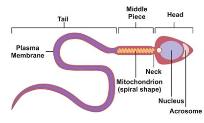
Sperm cell diagram with labels
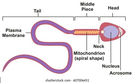
Draw the diagram of the human sperm and label its parts class 12 ... Complete step by step answer: - The human sperm can be divided into the head, the neck, the middle piece, and the tail. - The entire body is enveloped by a plasma membrane. - The head contains an elongated haploid nucleus that encloses the genetic material. The anterior end of the head is covered with a cap- like structure known as the acrosome. Sperm Cell, Egg Cell Diagram Label Worksheets (Differentiated) Three excellently differentiated worksheets. Engaging activity where pupils have to label the different parts of the male and femal gametes. Very well structured and scaffolded according to ability (from SEN to high ability). Excellent for visual learners. Compatible with all biology exam boards (including AQA, Edexcel, OCR). Sperm Cell - The Definitive Guide | Biology Dictionary The sperm cell diagram below shows multiflagellate fern cells. Sperm cells from the fern plant. Most motile spermatozoa have flagella to help them swim through fluids - the seminal fluid produced by males and the mucus membranes of the female reproductive tract. Flagellum movement requires a consistent energy source.
Sperm cell diagram with labels. Animal Cells: Labelled Diagram, Definitions, and Structure Plant cells have chloroplasts to synthesize their own food. Absent: Plasma Membrane: Cell wall and a cell membrane: Only cell membrane: Flagella: Present in some cells (e.g. sperm of bryophytes and pteridophytes, cycads and Ginkgo) Present in some cells ( e.g. mammalian sperm cells) Cilia: Most plant cells do not contain cilia. Present: Lysosomes Structure of Human Sperm: Check Types of Sperm - Embibe Explain the Structure of Human Sperm with Labelled Diagram Fig: Structure of a sperm cell Learn Exam Concepts on Embibe What is the Structure of Sperm? Human sperm is a microscopic structure whose shape is like a tadpole. It has flagella which make it motile. Its diameter is \ (2 - 5 {\rm { \mu m}},\) and its length is \ (60 {\rm { \mu m}}.\) Spermatozoa Development - Embryology This page introduces spermatogenesis the development of spermatozoa, the male haploid gamete cell. In humans at puberty, spermatozoa are produced by spermatogonia meiosis in the seminiferous tubules of the testis (male gonad). A second process of spermiogenesis leads to change in cellular organisation and shape before release into the central lumen of the seminiferous tubule. Draw the diagram of human sperm and label its parts. Write few lines ... Draw the diagram of human sperm and label its parts. Write few lines about it. Medium Solution Verified by Toppr The sperm cells are the haploid gametes which are produced in the male. There are different parts of the sperm cell. (a) Acrosome: This structure contains enzymes used for penetrating the female egg.
Labeled Sperm Cell Pictures, Images and Stock Photos Browse 15 labeled sperm cell stock photos and images available, or start a new search to explore more stock photos and images. Newest results Receptionist labeling sample in a laboratory Prostate labeled vector illustration. Educational male anatomy... Cell potency. From Totipotent to Pluripotent, Multipotent, and... Diagram and label sperm cell - Quizlet Only $2.99/month Diagram and label sperm cell STUDY Learn Flashcards Write Spell Test PLAY Match Gravity Created by Ike_SandersonTEACHER Terms in this set (4) Midsection of sperm contains mitochondria Sperm nucleus Contains haploid chromosomes Acrosome A vesicle at the tip of a sperm cell that helps the sperm penetrate the egg Flagellum Testes: Anatomy and Function, Diagram, Conditions, and Health Tips The epididymis stores sperm cells until they're mature and ready for ejaculation. ... Explore the interactive 3-D diagram below to learn more about the testes. ... (2015). "Off-label" usage ... Draw a labelled diagram of sperm. - Byju's Describe the various steps involved in the sexual reproduction in animals. Draw labelled diagrams to show the fertilisation of an ovum (or egg) by a sperm to form a zygote. Biology. Science for Tenth Class - Part III - Biology. Standard X.
Sperm Cell Labeled Diagram Stock Vector (Royalty Free) 200461103 ... Frequently used Trendsetter We're seeing significant engagement with this asset. Item ID: 200461103 Sperm Cell Labeled Diagram Formats EPS 6733 × 3563 pixels • 22.4 × 11.9 in • DPI 300 • JPG Contributor j joshya Similar images See all Assets from the same collection See all Similar video clips Sperm Cell Labeled Diagram Royalty Free Cliparts, Vectors, And Stock ... In Vitro Fertilisation Labeled Diagram Illustartion showing the structure of a sperm cell structure of the sperm in a section, male sperm cell closeup on white background Motor Neuron Disease beautiful design of sperms in the liquid Sperms and egg icon on origami paper speech bubble or web banner, prints. Vector illustration Sperm Diagram Stock Photos, Pictures & Royalty-Free Images - iStock Human Sperm cell Anatomy Male Doctor showing the male reproductive system. Mitosis and Meiosis Meiosis is a cell division in sexually reproducing organisms for produce the gametes. Meiotic phases: Prophase, Metaphase, Anaphase, and Telophase. Diagram of cell division Process. structure of a sperm cell Prostate gland Male reproductive system. labelled diagrams - the sperm cell labelled diagrams - the sperm cell to the right is a detailed 2D diagram of the sperm cell. there are many parts of a sperm cell. it is extremely small compared to the female egg.
Spermatogenesis Diagram & Function | What is the Process of Sperm ... Beneath the Sertoli cells are the spermatogonia, which are germ cells that will go through mitosis and ultimately create sperm. In humans, each day, roughly 25 million spermatogonia divide, and...
Sperm Cells Definition, Function, Structure, Adaptations & Microscopy The head of the sperm measures 2.5 to 3.5 um in diameter and 4.0 to 5.5 um in length (um=micrometers). This results in a 1.50 to 1.70 length to width ratio They have a well-developed acrosome that covers 40 to 70 percent of the oval shaped head A slim middle section (body) that is approximately the same length as the head
International GCSE Biology - Edexcel May 23, 2013 · 1 The diagram shows part of a food web in an oak forest. (a) Use the information in the food web to complete the statements in the table. The first one has been done for you. (4) Statement Number the number of animals is 8 the number of producers is the number of herbivores is the number of secondary consumers is the number of food chains is deer
What's the Function of a Sperm Cell? - Definition & Structure A spermatozoon, in plural spermatozoa, or sperm cell is the male reproductive cell that is expelled along with the seminal fluid or semen when a man ejaculates. In humans, spermatozoa determine the gender of the baby-to-be, which means that they can carry either the X or the Y chromosome.
PDF Pig Sperm Cell Diagram Labeled Pdf Free Download Pig Sperm Cell Diagram Labeled Pdf Free Download Author: hotels.propertyweek.com Subject: Pig Sperm Cell Diagram Labeled Keywords: Pig Sperm Cell Diagram Labeled, pdf, free, download, book, ebook, books, ebooks Created Date: 6/19/2022 9:44:59 PM
Draw a labeled diagram of sperm. - SaralStudy Q:-With a neat diagram explain the 7-celled, 8-nucleate nature of the female gametophyte. Q:-What is oogenesis? Give a brief account of oogenesis. Q:-What is DNA fingerprinting? Mention its application. Q:-With a neat, labelled diagram, describe the parts of a typical angiosperm ovule. Q:-What is triple fusion? Where and how does it take place?
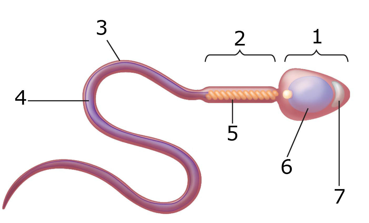
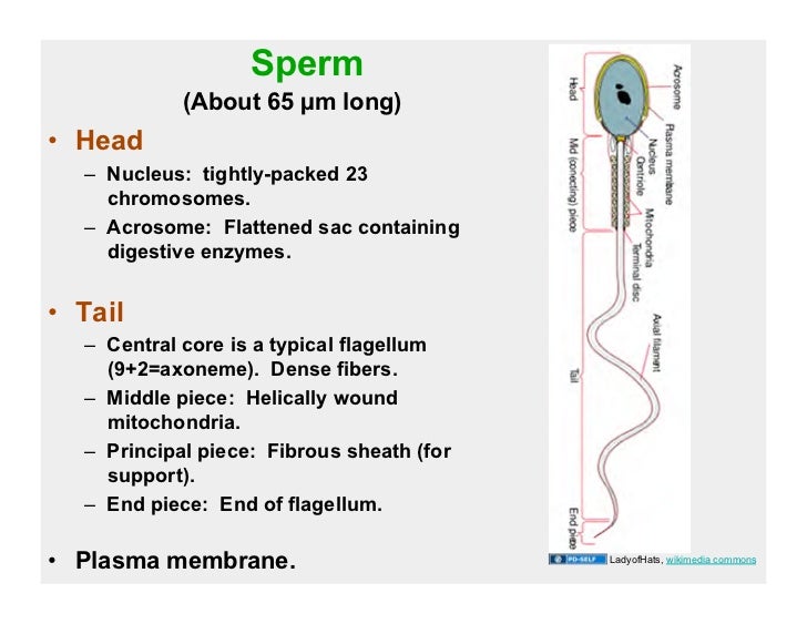
An overview of sperm anatomy | Legacy Sperm is the male sex cell, also known as a gamete. Measuring approximately 0.05 millimeter (0.002 inch) long, sperm cells are made up of a few distinct parts: the tail, made up of protein fibers, which helps it "swim" toward the egg the midpiece, or body, which contains mitochondria to power the sperm's movement
Sperm Cells Images | Free Vectors, Stock Photos & PSD Find & Download Free Graphic Resources for Sperm Cells. 500+ Vectors, Stock Photos & PSD files. Free for commercial use High Quality Images ... Diagram showing human sex cells on white background. brgfx. 8. Like. Collect. Save. In vitro fertilization flat elements. macrovector. 22. Like. Collect. Save. In vitro fertilization concept ...
Draw a diagram of the microscopic structure of human sperm. Label the ... The above diagram is of the sperm cell. (a) Acrosome: It contains enzymes used for penetrating the female egg. (b) Nucleus: Contains the genetic material that the sperm has to pass on, a haploid genome because it contains only one copy of each chromosome.
Sperm Diagram Stock Illustrations - 415 Sperm Diagram Stock ... Download 415 Sperm Diagram Stock Illustrations, Vectors & Clipart for FREE or amazingly low rates! ... Ocean depth zones infographic, vector illustration labeled diagram. Oceanography science educational graphic information. Depth at which sperm whales live and. ... Blue sperm cell vector illustration. 3d fertilisation isolated.
Biology: Chapter 10 Assignment Flashcards - Quizlet Fill in the Punnett square below to illustrate the possible genotypes of the offspring for the A gene. (Put alleles inherited from the mother on the top of the Punnett square and alleles inherited from the father along the left side of the Punnett square. Labels may be used more than once or not at all.)
Structure and parts of a sperm cell - inviTRA Structure and parts of a sperm cell 0 This labelled diagram shows the structure of a sperm cellin detail, which has the following parts: Head With its spheric shape, it consists of a large nucleus, which at the same time contains an acrosome. The nucleus contains the genetic information and 23 chromosomes.
Sperm Cell - The Definitive Guide | Biology Dictionary The sperm cell diagram below shows multiflagellate fern cells. Sperm cells from the fern plant. Most motile spermatozoa have flagella to help them swim through fluids - the seminal fluid produced by males and the mucus membranes of the female reproductive tract. Flagellum movement requires a consistent energy source.
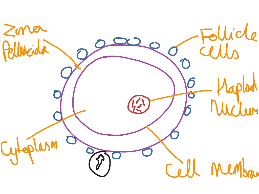
Sperm Cell, Egg Cell Diagram Label Worksheets (Differentiated) Three excellently differentiated worksheets. Engaging activity where pupils have to label the different parts of the male and femal gametes. Very well structured and scaffolded according to ability (from SEN to high ability). Excellent for visual learners. Compatible with all biology exam boards (including AQA, Edexcel, OCR).
Draw the diagram of the human sperm and label its parts class 12 ... Complete step by step answer: - The human sperm can be divided into the head, the neck, the middle piece, and the tail. - The entire body is enveloped by a plasma membrane. - The head contains an elongated haploid nucleus that encloses the genetic material. The anterior end of the head is covered with a cap- like structure known as the acrosome.




Post a Comment for "39 sperm cell diagram with labels"